Dr. Varshaa Hardas
Star Imaging And Research Centre
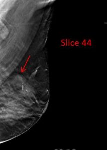
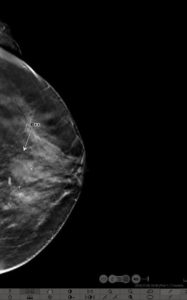
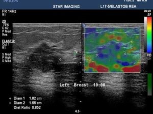
39yr old lady with history of lump in the left breast UIQ, No previous imaging done. No positive family history for CA Breast/Ovary. Pre-Menopausal Status.
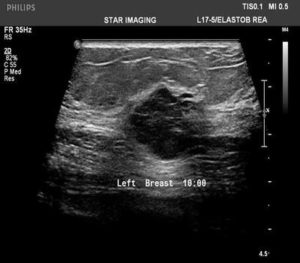
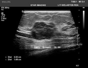
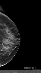
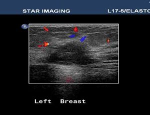
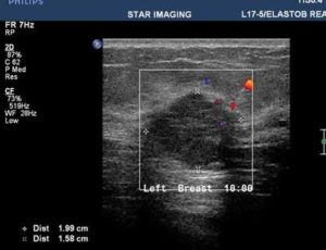
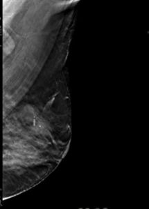
Mammography- Fairly well circumscribed predominantly sold lesion in the left breast UIQ with partially obscured margins. No spiculations. No associated microcalcifications noted. Sonography: Well defined hypoechoiec lesion with smooth margins.
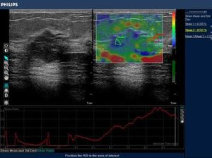
Low-grade Mucinous Carcinoma ER?PR- +ve HER-2 NEU -VE