Dr. Geetha priya, Dr. Priyadharshini,Dr.Sathyasree Dr.Bagyam Raghavan
Apollo Speciality Hospitals, Chennai.
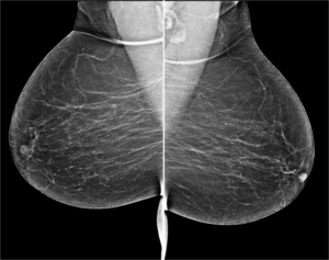
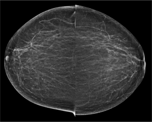
48 year old female came for routine mammographic screening
SKIN CALCIFICATIONS: Skin calcifications are single or clustered often they have a radiolucent centre with a calcific rim. They are always benign. These calcifications often simulate intraparenchymal calcifications. Skin or dermal calcifications are usually seen at the periphery of the breast, especially in the inferior, posterior, and medial aspects. Skin calcifications may develop from a degenerative metaplastic process. Skin calcifications are: Located peripherally in the breast Seen in the axilla, inframammary fold, or medial part of the breast Have a size similar to skin pores Located close to the skin surface on one view. When mammograms are compared, calcifications that maintain a fixed relationship to one another are suggestive of a dermal location. Magnification views may be used to demonstrate the lucent centers characteristic of skin calcifications. In some cases, a skin localization procedure may be needed to prove that the calcifications are in the skin.
Cranio caudal view: Focal cluster of punctate microcalcifications seen in left sub areolar region- Indeterminate. On Tomosynthesis: In the given slice of tomosynthesis the calcifications are clearly seen and also the skin pores are well made out in the same section which confirms that the calcifications are in the plane of skin. Hence the calcifications are benign. No further views/ management required.
Breast Imaging: The Requisites, 2nd edition, Ikeda D, Chapter-3. Mammographic Analysis of Breast Calcifications. 2010; ISBN-10: 0323051987